Myocardial Infarction
Myocardial Infarction
Myocardial Infarction is the term for an event commonly known as a “HEART ATTACK”. It occurs when blood stops flowing properly to part of the heart and the heart muscle is injured due to not receiving enough oxygen. Usually this is because one of the coronary arteries that supplies blood to the heart develops a blockage due to an unstable buildup of white blood cells, cholesterol and fat. The event is called “acute” if it is sudden and serious. Superimposed thrombus in Atherosclerotic coronary vessels is the most common cause (>90 percent cases) plaque rupture, plaque hemorrhage and super added spasm are additional factors. Non-Atherosclerotic Causes Include Coronary arteritis (collagen disease, Kawasaki disease). Coronary embolism from endocarditis, myxoma, LA thrombus Hypercoagulability and poly cythemia Reduced oxygen delivery due to hypotension,anaemia,carbon monoxide,hamoglobinopatheis. Increased O2, demand from thyrotoxicosis, LVH Anomalous orgin of left coronary from pulmonary artery. Prodrome None in 50 percent cases Unstable angina.
Signs and Symptoms in Myocardial Infarction
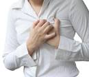
The onset of symptoms in myocardial infarction (MI) is usually gradual, over several minutes, and rarely instantaneous.Chest pain is the most common symptom of acute MI and is often described as a sensation of tightness, pressure, or squeezing. Chest pain due to ischemia (a lack of blood and hence oxygen supply) of the heart muscle is termed angina pectoris. Pain radiates most often to the left arm, but may also radiate to the lower jaw, neck, right arm, back, and epigastrium,where it may mimic heartburn. Levine’s sign, in which patients localize the chest pain by clenching their fists over their sternums, has classically been thought to be predictive of cardiac chest pain, although a prospective observational study showed it had a poor positive predictive value.

Chest Pain in Myocardial Infarction
Pain
Crushing, constricting, retrosternal pain exceeding 30 minutes; radiating to ulnar aspect of left arm, interscapular area, neck, jaw and right arm, not relieved by nitroglycerine. Sweating, vomiting, epigastric distress Syncope, sudden death Acute pulmonary edema (LVF) Weakness and confusion (especially in elderly) Shortness of breath (dyspnoea) occurs when the damage to the heart limits the output of the left ventricle, causing left ventricular failure and consequent pulmonary edema. Other symptoms include diaphoresis (an excessive form of sweating), weakness, light-headedness, nausea, vomiting, and palpitations. These symptoms are likely induced by a massive surge of catecholamines from the sympathetic nervous system which occurs in response to pain and the hemodynamic abnormalities that result from cardiac dysfunction. Loss of consciousness (due to inadequate cerebral perfusion and cardiogenic shock) and sudden death (frequently due to the development of ventricular fibrillation) can occur in MI. Variations Pain may be absent in some due to concomitant neuropathy (esp.diabetes) Right heart failure of sudden onsent due to RV infarction Parasympathetic over activity with nausea and vomiting, bloating in inferior infarction.
Diagnosis of Myocardial Infarction
WHO criteria formulated in 1979 have classically been used to diagnose MI; a patient is diagnosed with MI if two (probable) or three (definite) of the following criteria are satisfied: Clinical history of ischaemic type chest pain lasting for more than 20 minutes Changes in serial ECG tracings Rise and fall of serum cardiac biomarkers.
Physical Examination findings Anxious, diaphoresis, restless, Evidence of CHF or pulmonary congestion (RV and LV infarction) Low grade fever Blood pressure, a drop from previous level. S2 when present indicates poor prognosis Systolic murmur due to (a) Papillary muscle dysfunction (b) Ruptured I.V. septum Pericardial rub.
Acute Myocardial Infraction : Auscultatory signs Sounds 1st soft, 2nd paradoxical split 3rd gallop Murmur Systolic apical Pericardial rub.
ECG Changes in Different Leads in Relation to Location of Infarction Frequent complex VPBs, widespread Q waves; A-V and I-V conduction disturbances.

myocardial infarction

|
Leads |
Location |
| V1-2V1-4V3-4V5-V6 V5-6, I, aVL V3-6, I, aVL I, AVL II, III,aVF | SeptalAnteroseptalAnteriorApical Lateral Antero lateral High lateral Inferior |
X-Ray findings
Cardiomegaly.A chest radiograph and routine blood tests may indicate complications or precipitating causes, and are often performed upon arrival to an emergency department.
CPK > 2000 U / ml
Echo findings
New regional wall motion abnormalities on an echocardiogram are also suggestive of an MI. Imparied LV function (ejection fraction < 40 percent).stressechocardiography can confirm a diagnosis when a patient’s history, physical exam, ECG, and cardiac biomarkers suggest the likelihood of a problem. Regional wall motion abnormality Detection of complications, (a) Pericardial effusion, (b) MR,PMW, (c) LV clot, (d) VSD.
Thallium Study Perfusion defect (cold spot) Stannous Pyrophosphate Hot spot (useful after 24 hours) Antimyosin antibody imaging More specific but results will be available 24-28 hours post injection.
Clinical Utility of Hotspot Scanning Suspected infarct with negative enzymes (e.g. late presentation) Suspected infarct with equivocal enzymes (e.g. post-surgery, intramuscular injection) Suspected infarct with non-diagnostic ECG In stable patients whose symptoms have resolved by the time of evaluation, technetium (99mTc) sestamibi (i.e. a “MIBI scan”) or thallium-201 chloride can be used in nuclear medicine to visualize areas of reduced blood flow in conjunction with physiological or pharmacological stress.Thallium may also be used to determine viability of tissue, distinguishing whether nonfunctional myocardium is actually dead or merely in a state of hibernation or of being stunned.
Management of Myocardial Infarction
An MI requires immediate medical attention. Treatment attempts to save as much myocardium as possible and to prevent further complications, hence the phrase “time is muscle”.Oxygen, aspirin, and nitroglycerin may be administered. Morphine was classically used if nitroglycerin was not effective; however, it may increase mortality in the setting of NSTEMI.Reviews of high flow oxygen in myocardial infarction found increased mortality and infarct size, calling into question the recommendation about its routine use. Absolute bed rest, Aspirin 350 mg PO Injection Morphine 3 mg. I.V. every 15 minutes O2 inhalation I.V. streptokinase 1.5 mill. units / urokinase / rtPA / APSAC within 6 hours of pain or before appearonce of Qwaves.
Antiplatelet drug therapy such as aspirin and/or clopidogrel should be continued to reduce the risk of plaque rupture and recurrent MI. Aspirin is first-line, owing to its low cost and comparable efficacy, with clopidogrel reserved for patients intolerant of aspirin. The combination of clopidogrel and aspirin may further reduce risk of cardiovascular events, but the risk of hemorrhage is increased.
Sorbitrate 10 mg every 6 hours.
Beta blocker therapy such as metoprolol or carvedilol should be started.Betablockers metoprolol 5 mg IV (3 doess) or esmolol 30mg IV and nifedipine.These have been particularly beneficial in those who are high-risk such as those with left ventricular dysfunction and/or continuing cardiac ischaemia.β-Blockers decrease mortality and morbidity. They also improve symptoms of cardiac ischemia in NSTEMI.
ACE inhibitors to improve remodeling.ACE inhibitor therapy should be commenced 24–48 hours after MI in those who are hemodynamically stable, particularly with a history of MI, diabetes mellitus, hypertension, anterior location of infarct (as assessed by ECG), and/or evidence of left ventricular dysfunction. ACE inhibitors reduce mortality, the development of heart failure, and decrease ventricular remodelling.
Statin therapy has been shown to reduce mortality and morbidity.The effects of statins may be more than their LDL lowering effects. The general consensus is that statins have plaque stabilization and multiple other (“pleiotropic”) effects that may prevent myocardial infarction in addition to their effects on blood lipids.
The aldosterone antagonist agent eplerenone has been shown to further reduce risk of cardiovascular death after MI in patients with heart failure and left ventricular dysfunction, when used in conjunction with standard therapies above.Spironolactone, another option, is sometimes preferable to eplerenone due to cost. Evidence supports the consumption of polyunsaturated fats instead of saturated fats as a measure of decreasing coronary heart disease.In high-risk people, no clear-cut decrease in potentially fatal arrhythmias occurs due to omega-3 fatty acids.And they may increase risk in some groups.
Prophylactic heparin 5000 IU SC twice daily/fragnin.Giving heparin to people with heart conditions like unstable angina and some forms of heart attacks reduces the risk of having another heart attack. However, heparin also increases the chance of minor bleeding. Liquid diet and stool softners.
Percutaneous coronary intervention (PCI) is the treatment of choice for STEMI if it can be performed in a timely manner.If PCI cannot be performed within 90 to 120 minutes then fibrinolysis, preferably within 30 minutes, is recommended.If after fibrinolysis, significant cardiogenic shock, continued severe chest pain, or less than a 50% improvement in ST elevation after 90 minutes occurs, then rescue PCI is indicated emergently.After PCI, people are generally placed on dual antiplatelet therapy for at least a year (which is generally aspirin and clopidogrel). Balloon angioplasty to be considered if infarction progressive or causing hemodynamic compromise.
Coronary artery interventions Balloon angioplasty Balloon angioplasty and stenting Balloon angioplasty and brachytherapy Balloon angioplasty with VGEF/FGF Rotablator Directional atherectomy Noncoronary interventions Transmyocardial revascularization Cardio myoplasty LV assist device Cardaic transplant
Complications in Myocardial Infarction
Early Arrhythmias
Burst (Rupture) Vent.septum,
cardiac Collapse (Shock)
Dysfunction of papillary muscle
Embolism
Failure : LV or congestive Late : ASD Aneurysm (ventricular) Shoulder hand syndrome Dressler’s syndrome.
Bradycardia if symptomatic Atropine, isoprenaline, steroid, if not – pacing Tachy arrhythmias Ventricular fibrillation – defibrillator If VF is of late onset – recurrence is great and is a poor prognostic feature Multiform VPBs, R on T phenomena -Xylocaine infusion Heart Block Anterior infarction – Pacing Inferior infarction – Steroids and atropine ; if not. temporary pacing.
Heart Failure R.V.Failure : Fluids, intropic agents L.V.failure : Diuretics, vasodilators, inotropic agents, balloon counter pulsation (digoxin contra-indicated unless tachyarhytmia causes heart failure) Aneurysm Diagnosed from persistend ST elevation beyond 8 weeks. (a) Mural thrombus in anterior MI need IV heparin / fragmin. (b) Myocardial rupture, papillary mucle rupture, pericarditis.
Myocardial infarction due to coronary lesion other than atheroma.
Anomalous origin of coronary artery.
Bulge (aneurysm) of cor. artery
Collagen disease : SLE, PAN
Dissecting aneurysm of cor. artery
Embolism (e.g. SBE)
Fistula of cor. artery
Fibrosis following irradiation
Prognosis in Myocardial Infarction
Half of myocardial infarction deaths occur due to fatal arrhythmia (VF) occurring before the patient reaches hospital. Hence, effective C.P. resuscitation and prophylactic xylocaine infusion can prevent some of these deaths. Inhospital mortality is predicted by extent of infarction and its effect on L.V. function. Patients in Killip III and IV have high (40-80 percent) mortality. Late mortality is 5% per year for first 5 years and then less. This can be reduced by prophylactic betablocker, therapy for minimum 5 years.
Poor Prognosis (1) Cardiogenic shock, (2) Old age, (3) Previous infarct, (4) Post-infarct angina and / or persistent tachycardia.
Advice on Discharge
Limited stress test to evaluate arrhythmias and exercise tolerance so that a suitable work programme could be prescribed. Abstinence from smoking and tobacco in any form for rest of life. Coronary bypass surgery for suitable candidates. Folic acid 1mg / kg, B5 and B12 to reduce homocystine. Aspirin 75 mg daily or ticlopidine 250 mg PO to reduce platelet aggregation. Antioxidants (Vit A, E and C) ; Betablocker ACE inhibitor (in large infraction, CHF, low EF)
Cigarette Smoking and Cardiovascular Sequelae
Increased platelet adhesion / aggregation / whole blood viscosity. Increased heart rate, increased catecholamine sensitivity / release. Increase carboxyhamemoblobin level, increased haematocrit Decreased level of HDL-cholesterol/vascular compliance Decreased threshold to ventricular fibrillation Endothelial dysfunction


